IMPORTANT HEALTH/MEDICAL NUMBERS IN HUMANS

IMPORTANT MEDICAL NUMBERS IN THE LIFE OF EVERY HUMAN BEING*

  1. Blood pressure: 120 / 80
  2. Pulse: 70 – 100
  3. Temperature: 36.8 – 37
  4. Respiration: 12-16
  5. Hemoglobin: Males (13.50-18)
    Females ( 11.50 – 16 )
  6. Cholesterol: 130 – 200
  7. Potassium: 3.50 – 5
  8. Sodium: 135 – 145
  9. Triglycerides: 220
  10. The amount of blood in the body:
    Pcv 30-40%
  11. Sugar: for Children (70-130)
    Adults: 70 – 115
  12. Iron: 8-15 mg
  13. White blood cells: 4000 – 11000
  14. Platelets: 150,000 – 400,000
  15. Red blood cells: 4.50 – 6 million..
  16. Calcium: 8.6 – 10.3 mg/dL
  17. Vitamin D3: 20 – 50 ng/ml (nanograms per milliliter)
  18. Vitamin B12: 200 – 900 pg/ml Tips for those who have reached Over:
    the 40years
    the 50
    the 60 First tip:
    Always drink water even if you don’t feel thirsty or need it… the biggest health problems and most of them are from the lack of water in the body. 2 litres Minimum per day (24 hours)

Second tip:
Play sports even when you are at the top of your preoccupation…the body must be moved, even if only by walking…or swimming…or any kind of sports. 🚶 Walking is Good for a Start… 👌

Third Tip:
Reduce food…

Leave excessive food cravings… because it never brings good. Don’t deprive yourself, but reduce the quantities. Use more Of Protein, Carbohydrates based Foods.

Fourth tip
As much as possible, do not use the Car unless absolutely necessary… Try to reach on your Feet for what you want ( grocery, visiting someone…) or any goal. Climb Stairs, than use Elevator, Escalator

Fifth tip
let go of ANGER…
let go of Anger…
let go of anger…
Let go of Worry…. try to overlook things…

Do not involve yourself in situations of Disturbance… they all diminish health and take away the splendor of the soul. Choose a babysitter you feel comfortable with. Talk to People who are Positive and Listen 👂

Sixth tip
As it is said..leave your Money in the Sun.. and sit in the Shade.. don’t limit yourself and those around you… money was made to live By it, not to live For it.

Seventh tip
Don’t make yourself feel Sorry for anyone, nor on something you could not Achieve,
Nor anything that you could not Own.
Ignore it, Forget it;

Eighth tip
Humility.. then humility.. for Money, Prestige, Power and Influence… they are all things that are corrupted by Arrogance and arrogance..
Humility is what brings people Closer to you with Love. ☺

Ninth tip
If your hair turns Grey, this does not mean the End of Life. It is proof that a Better life has Begun. 🙋 Be Optimistic, live with Remembrance, Travel, Enjoy yourself. Make Memories!

It may help someone when you share it👀
TO ALL ELDERS IN THE HOUSE, KINDLY FOLLOW ME UP. IT IS VERY NECESSARY TO FOLLOW THIS INSTRUCTION STEP BY STEP.

HEALTH HINTS FOR MY ELDERS


However busy you are, observe all these to remain healthy:
::::::::::::::::::::::::::::::::::::
Drink less milk in your tea. Instead, add lemon or lime juice.

In the day time, drink more water; but night time, drink less. 

In the day don’t drink more than 2 cups of coffee, Advisable To Stop Completely too.

Eat less oily foods. 

Best sleeping times are between 10pm to 6am.

In the evening, eat little or nothing after 5 or 6pm. 
~~~~~~~~~~~~~
Don't take medicines with cold water but with warm, and take your medicines half an hour before going to bed. Never take medicines and lie down immediately. 

As you aged further , stop drinking chilled water but drink only water at room temperature

Try to sleep for at least 8 hours per day.

Having a nap for an hour and a half between noon and 3pm, to relieve stress and keep younger and not age easily.

Once your mobile phone battery is left with only one bar, don't make calls anymore, because the dangerous radiation and waves are one many times higher than a fully charged battery.

Use your left ear to answer calls, right ear will directly hurt your brain. 😳 Better still to use earphones to answer calls.

Two things to check as often as you can:
(1) Your blood pressure
(2) Your blood sugar.

Six things to reduce to the minimum on your foods:
(1) Salt
(2) Sugar
(3) Preserved meat and foods
(4) Red meat especially roasted
(5) Dairy products
(6) Starchy products

Four things to increase in your foods:
(1) Greens/vegetables 
(2) Beans 
(3) Fruits 
(4) Nuts 

Three things you need to forget:
(1) Your age 😮
(2) Your past 🤔
(3) Your worries/grievances 👍🏽

Four things you must have, no matter how weak or how strong you are: 
(1) Friends who truly love you
(2) Caring family
(3) Positive thoughts
(4) A warm home.

Seven things you need to do to stay healthy:
(1) Singing
(2) Dancing
(3) Fasting
(4) Smiling/laughing
(5) Trek/exercise
(6) Have sex often with your love
(7) Reduce your weight.

Six things you don't have to do: 
(1) Don't wait till you are hungry to eat
(2) Don't wait till you are thirsty to drink
(3) Don't wait till you are sleepy to sleep
(4) Don't wait till you feel tired to rest
(5) Don't wait till you get sick to go for medical check-ups otherwise you will only regret later in life
(6) Don’t wait till you have problem before you pray to your God.

One thing you must do after reading these health tips:

(1) Forward this to your loved ones and friends, and as you do so, may God bless U.

While go about your normal business please let’s remember to always check our body to know how fit you are. Health is wealth.

MEDICAL FITNES

       HIGH BP
      ----------

120/80 — Normal
130/85 –Normal (Control)
140/90 — High

150/95 — V.High

       PULSE
      --------

72 per minute (standard)
60 — 80 p.m. (Normal)

40 — 180 p.m.(abnormal)

      TEMPERATURE
      -----------------

98.4 F (Normal)
99.0 F Above (Fever)

Please help your Relatives, Friends by sharing this information….

Heart Attacks- – –
Drinking Warm
Water:

This is a very good article. Not only about the warm water after your meal,
but about Heart Attack’s . The Chinese and Japanese drink hot tea with their
meals, not cold water, maybe it is time we adopt
their drinking habit while
eating. For those who like to drink cold water, this
article is applicable to
you. It is very Harmful to have Cold Drink/Water during a meal. Because,
the cold water will solidify the oily stuff that you
have just consumed. It will
slow down the digestion. Once this ‘sludge’
reacts with the acid, it will break down and be
absorbed by the intestine faster than the solid food. It will line the intestine. Very soon, this will turn into fats
and lead to cancer . It is best to drink hot soup
or warm water after a meal.

French fries and Burgers
are the biggest enemy of heart health. A coke after that gives more power to
this demon. Avoid them for
your Heart’s & Health.

Drink one glass of warm water just when you are about to go to bed to avoid clotting of the blood at night to avoid heart attacks or strokes.

A cardiologist says if everyone who reads this
message sends it to 10 people, you can be sure that we’ll save at least
one life. …

So, please be a true friend and send this article to people you care about.

Must read 👆👌

Cheers And Enjoy life
Know your genotype before you say yes to that handsome guy or to that beautiful lady whom you wish to spend the rest of your life with…
Genotype & It’s Appropriate Suitor:
AA + AA = Excellent
AA + AS = Good
AA + SS = Fair
AS + AS = Bad
AS + SS = Very Bad
SS + SS = Extremely Bad (In fact, don’t try it)

SickleCellAwareness

💉BLOOD GROUP COMPATIBILITY 💉

What’s Your Type and how common is it?

O+ 1 in 3 37.4%
(Most common)

A+ 1 in 3 35.7%

B+ 1 in 12 8.5%

AB+ 1 in 29 3.4%

O- 1 in 15 6.6%

A- 1 in 16 6.3%

B- 1 in 67 1.5%

AB- 1 in 167 .6%
(Rarest)

Compatible Blood Types

O- can receive O-

O+ can receive O+, O-

A- can receive A-, O-

A+ can receive A+, A-, O+, O-

B- can receive B-, O-

B+ can receive B+, B-, O+, O-

AB- can receive AB-, B-, A-, O-

AB+ can receive AB+, AB-, B+, B-, A+, A-, O+, O-b

This is an important msg which can save a life! A life could be saved….
: Your Blood group also speaks about you.
🅰️(+) : Good leadership.
🅰️(-) : Hardworking.
🅱️(+) : Can Sacrifice for others and very ambitious, tolerance.
🅱️(-) : Non flexible, Selfish & Sadistic.
🅾️(+) : Born to help.
🅾️(-) : Narrow minded.
🆎(+) : Very difficult to understand.
🆎(-) : Sharp & Intelligent.

What is ur blood group ?
Try….It….share the Fantastic information..
EFFECT OF WATER
💐 We Know Water is
important but never
knew about the
Special Times one
has to drink it.. !!

   Did you  ???  

💦 Drinking Water at the
Right Time ⏰
Maximizes its
effectiveness on the
Human Body;

   1⃣  1 Glass of Water 
          after waking up -
         🕕⛅ helps to 
          activate internal 
          organs..

   2⃣  1 Glass of Water 
          30 Minutes  🕧 
          before a Meal - 
          helps digestion..

   3⃣ 1 Glass of Water 
          before taking a 
          Bath 🚿 - helps 
          lower your blood 
          pressure.

   4⃣ 1 Glass of Water 
          before going to 
          Bed - 🕙 avoids 
          Stroke  or Heart 
          Attack.

  'When someone 
   shares something of 
   value with you and 
   you benefit from  it, 
   You have a moral 
   obligation to share it 
   with others.

How to detect early signs of heart disease in the eyes

Early Signs of Heart Diseases Appear in the Eyes

According to experts, Eye doctors may be able to detect signs of heart disease during a comprehensive eye exam. A new study finds that people with heart disease tend to have retinas marked by evidence of eye stroke.

Eye strokes happen when the eye is deprived of blood flow and oxygen, causing cells to die. This creates a mark, called a retinal ischemic perivascular lesion. These marks can be spotted when eye doctors run optical coherence tomography, or OCT is order to take a closer look at the retina.

OCT scans of the retina are valuable ways to detect disease and dysfunction in all parts of the body — not just the eyes. Eye scans can detect signs of Alzheimer’s, Parkinson’s and other underlying health conditions.

How eye exams can detect heart disease

The eye is the only place in the body where a doctor can see the live action of blood vessels, nerves and connecting tissue without relying on an invasive procedure. That’s why eye doctors are often the first to detect health conditions including high blood pressure, high cholesterol, stroke and more.

While the marks left behind by eye strokes may be present in low numbers in healthy people, those with heart disease tend to have a far greater number. Researchers arrived at these results by reviewing the medical records of 84 people with heart disease and 76 healthy people, all of whom had received a retinal OCT scan.

Visual field testing

What is visual field testing?

As you focus on the words in this article, how much can you see out of the corners of your eyes? Can you tell what’s happening in your surroundings?

Your visual field is how wide of an area your eye can see when you focus on a central point. Visual field testing is one way your ophthalmologist measures how much vision you have in either eye, and how much vision loss may have occurred over time.

Visual field testing can detect blind spots

A visual field test can determine if you have blind spots (called scotoma) in your vision and where they are. A scotoma’s size and shape can show how eye disease or a brain disorder is affecting your vision. For example, if you have glaucoma, this test helps to show any possible side (peripheral) vision loss from this disease.

Ophthalmologists also use visual field tests to assess how vision may be limited by eyelid problems such as ptosis and droopy eyelids.

Six types of visual field tests

1. Confrontation visual field test

A common way for your doctor to screen for any problems in your visual field is with a confrontation visual field test. You will be asked to look directly at an object in front of you, (such as the doctor’s nose) while one of your eyes is covered. Your doctor may hold up different numbers of fingers in areas of your peripheral (side) vision field and ask how many you see as you look at the target in front of you.

2. Automated static perimetry test

To check for a suspected eye problem or monitor the progress of an eye disease, your ophthalmologist will rely on more specific tests to measure how you see objects in your field of vision. The automated static perimetry test is used for this purpose. It helps create a more detailed map of where you can and can’t see.

To do this test, you will look into the center of a bowl-shaped instrument called a perimeter. The eye not being tested will be covered with a patch. The testing eye will have your lens prescription placed in front of it to make sure you are seeing as well as possible.

A patient sits at a perimeter machine taking a visual field exam.

You will be asked to keep looking at a center target throughout the test. Small, dim lights will begin to appear in different places throughout the bowl, and you will press a button whenever you see a light. The machine tracks which lights you did not see.

You may blink normally during the test. You may also pause the test if you feel you need to take a break for a moment.

Because you are looking straight ahead during the test, your doctor can tell which lights you see outside of your central area of vision. Since glaucoma affects peripheral vision, this test helps show if there is vision loss outside of your central visual field.

This is called a “static” test because the lights do not move across the screen, but blink at each location with differing amounts of brightness. This allows the machine to find the dimmest light you can see at each location in your peripheral vision.

The machine will show some lights that are too dim for you to see. This is done deliberately to find what is called the “visual threshold” of each location, meaning the brightness that you have trouble seeing half the time. You may be concerned because you can’t see every light. Rest assured that this is how the test is supposed to work.

Normal visual field test results

A printout of normal visual field results.

3. Kinetic visual field test

In some cases, you may have a test called kinetic visual field testing. While it is similar to the perimetry testing process described above, the kinetic test uses moving light targets instead of blinking lights.

4. Frequency doubling perimetry

Another way your ophthalmologist can assess loss of vision is using something called frequency doubling perimetry. It uses an optical illusion to check for damage to vision. Vertical bars (usually black and white) appear on the perimeter screen. These bars will flicker at varying rates. If you are not able to see the vertical bars at certain times during the test, it could show vision loss in certain parts of your visual field.

5. Electroretinography

To check for visual field loss from certain retina conditions, your ophthalmologist may also use electroretinography (ERG). This test measures the electrical signals of light-sensitive cells in the retina called photoreceptors as well as other cells. To do this test, your eyes are dilated and you will also be given numbing eye drops. Your eyes are held open with instrument called a speculum. A tiny device called an electrode is placed on your cornea. You will look into a bowl-shaped machine at flashing or varying patterns of light. The electrode measures your eye’s electrical activity in response to the light.

6. Amsler grid: A basic visual field test for central vision

People who have age-related macular degeneration (AMD) are familiar with one very basic type of visual field test: the Amsler grid. It is a pattern of straight lines that makes a grid of many equal squares. You look at a dot in the middle of the grid and describe any areas that may appear wavy, blurry or blank.Amsler Grid, used at home daily to track vision changes in people with some eye conditions like AMD or toxoplasmosisAmsler Grid

The Amsler grid is commonly used at home by people with AMD. This test only measures the middle of the visual field, but is a simple yet helpful tool for monitoring vision changes.

How do you know if you need visual field testing?

Visual field testing is an important part of regular eye care for people who are at risk for vision loss from disease and other problems.

People with the following conditions should be monitored regularly by their ophthalmologist, who will determine how often visual field testing is needed:

  • Glaucoma
  • Multiple sclerosis
  • Thyroid eye disease (Graves’ disease)
  • Pituitary gland disorders
  • Central nervous system problems (such as a tumor that may be pressing on visual parts of the brain)
  • Stroke
  • Long-term use of certain medications (such as Plaquenil, or hydroxychloroquine, which requires yearly visual field checkups)

People with diabetes and high blood pressure have a greater risk of developing blocked blood vessels in the optic nerve and retina. They may need visual field testing to monitor any effects of these conditions on their vision.

If your visual field is limited, your ability to drive may be in jeopardy. If you are concerned about vision loss or your ability to continue driving, talk with an eye care practitioner.

You can have your visual field testing at Eyeupdate Eye clinic, 01, Ajuwon bus stop, Akute-Ajuwon Road via Iju Ishaga.

How to prevent vision loss

Follow these simple guidelines for maintaining healthy eyes well into your golden years.

Your eyes are an important part of your health. You can do many things to keep them healthy and make sure you’re seeing your best. Follow these simple guidelines for maintaining healthy eyes well into your golden years.

Have a comprehensive dilated eye exam. You might think your vision is fine or that your eyes are healthy, but visiting your eye care professional for a comprehensive dilated eye exam is the only way to really be sure. When it comes to common vision problems, some people don’t realize they could see better with glasses or contact lenses. In addition, many common eye diseases, such as glaucoma, diabetic eye disease, and age-related macular degeneration, often have no warning signs. A dilated eye exam is the only way to detect these diseases in their early stages.

During a comprehensive dilated eye exam, your eye care professional places drops in your eyes to dilate, or widen, the pupil to allow more light to enter the eye—the same way an open door lets more light into a dark room. This process enables your eye care professional to get a good look at the back of the eyes and examine them for any signs of damage or disease. Your eye care professional is the only one who can determine if your eyes are healthy and if you’re seeing your best.

Maintain your blood sugar levels. 90% of blindness caused by diabetes is preventable. Ask your health care team to help you set and reach goals to manage your blood sugar, blood pressure, and cholesterol—also known as the ABCs of diabetes.

  • A1c: The goal set for many people is less than 7% for this blood test, but your doctor might set different goals for you.
  • Blood pressure: High blood pressure causes heart disease. The goal is less than 140/90 mmHg for most people, but your doctor might set different goals for you.
  • Cholesterol: LDL or “bad” cholesterol builds up and clogs your blood vessels. HDL or “good” cholesterol helps remove the “bad” cholesterol from your blood vessels. Ask what your cholesterol numbers should be.

Know your family’s eye health history. Talk to your family members about their eye health history. It’s important to know if anyone has been diagnosed with an eye disease or condition, since many are hereditary. This information will help to determine if you’re at higher risk for developing an eye disease or condition.

Eat right to protect your sight. You’ve heard that carrots are good for your eyes. But eating a diet rich in fruits and vegetables—particularly dark leafy greens, such as spinach, kale, or collard greens—is important for keeping your eyes healthy, too.i Research has also shown there are eye health benefits from eating fish high in omega-3 fatty acids, such as salmon, tuna, and halibut.

Maintain a healthy weight. Being overweight or obese increases your risk of developing diabetes and other systemic conditions, which can lead to vision loss, such as diabetic eye disease or glaucoma. If you’re having trouble maintaining a healthy weight, talk to your doctor.

Wear protective eyewear. Wear protective eyewear when playing sports or doing activities around the home. Protective eyewear includes safety glasses and goggles, safety shields, and eye guards specially designed to provide the correct protection for the activity in which you’re engaged. Most protective eyewear lenses are made of polycarbonate, which is 10 times stronger than other plastics. Many eye care providers sell protective eyewear, as do some sporting goods stores.

Quit smoking or never start. Smoking is as bad for your eyes as it is for the rest of your body. Research has linked smoking to an increased risk of developing age-related macular degeneration, cataract, and optic nerve damage, all of which can lead to blindness.ii, iii

Be cool and wear your shades. Sunglasses are a great fashion accessory, but their most important job is to protect your eyes from the sun’s ultraviolet rays. When purchasing sunglasses, look for ones that block out 99 to 100 percent of both UV-A and UV-B radiation.

Give your eyes a rest. If you spend a lot of time at the computer or focusing on any one thing, you sometimes forget to blink and your eyes can get fatigued. Try the 20-20-20 rule: Every 20 minutes, look away about 20 feet in front of you for 20 seconds. This short exercise can help reduce eyestrain.

Clean your hands and your contact lenses—properly. To avoid the risk of infection, always wash your hands thoroughly before putting in or taking out your contact lenses. Make sure to disinfect contact lenses as instructed and replace them as appropriate. Learn more about keeping your eyes healthy while wearing contact lenses and listen to a podcast on keeping your eyes safe.

Practice workplace eye safety. Employers are required to provide a safe work environment. When protective eyewear is required as a part of your job, make a habit of wearing the appropriate type at all times, and encourage your coworkers to do the same.

Eye care and eye safety tips

Your eyes are an important part of your health. Most people rely on their eyes to see and make sense of the world around them. But some eye diseases can lead to vision loss, so it is important to identify and treat eye diseases as early as possible. You should get your eyes checked as often as your health care provider recommends it, or if you have any new vision problems. And just as it is important to keep your body healthy, you also need to keep your eyes healthy.

Eye Care Tips

There are things you can do to help keep your eyes healthy and make sure you are seeing your best:

  • Eat a healthy, balanced diet. Your diet should include plenty or fruits and vegetables, especially deep yellow and green leafy vegetables. Eating fish high in omega-3 fatty acids, such as salmon, tuna, and halibut can also help your eyes.
  • Maintain a healthy weight. Being overweight or having obesity increases your risk of developing diabetes. Having diabetes puts you at higher risk of getting diabetic retinopathy or glaucoma.
  • Get regular exercise. Exercise may help to prevent or control diabetes, high blood pressure, and high cholesterol. These diseases can lead to some eye or vision problems. So if you exercise regularly, you can lower your risk of getting these eye and vision problems.
  • Wear sunglasses. Sun exposure can damage your eyes and raise your risk of cataracts and age-related macular degeneration. Protect your eyes by using sunglasses that block out 99 to 100% of both UV-A and UV-B radiation.
  • Wear protective eye wear. To prevent eye injuries, you need eye protection when playing certain sports, working in jobs such as factory work and construction, and doing repairs or projects in your home.
  • Avoid smoking. Smoking increases the risk of developing age-related eye diseases such as macular degeneration and cataracts and can damage the optic nerve.
  • Know your family medical history. Some eye diseases are inherited, so it is important to find out whether anyone in your family has had them. This can help you determine if you are at higher risk of developing an eye disease.
  • Know your other risk factors. As you get older, you are at higher risk of developing age-related eye diseases and conditions. It is important to know you risk factors because you may be able to lower your risk by changing some behaviors.
  • If you wear contacts, take steps to prevent eye infections. Wash your hands well before you put in or take out your contact lenses. Also follow the instructions on how to properly clean them, and replace them when needed.
  • Give your eyes a rest. If you spend a lot of time using a computer, you can forget to blink your eyes and your eyes can get tired. To reduce eyestrain, try the 20-20-20 rule: Every 20 minutes, look away about 20 feet in front of you for 20 seconds.

Eye Tests and Exams

Everyone needs to have their eyesight tested to check for vision and eye problems. Children usually have vision screening in school or at their health care provider’s office during a checkup. Adults may also get vision screenings during their checkups. But many adults need more than a vision screening. They need a comprehensive dilated eye exam.

Getting comprehensive dilated eye exams is especially important because some eye diseases may not have warning signs. The exams are the only way to detect these diseases in their early stages, when they are easier to treat.

The exam includes several tests:

  • A visual field test to measure your side (peripheral) vision. A loss of peripheral vision may be a sign of glaucoma.
  • A visual acuity test, where you read an eye chart about 20 feet away, to check on how well you see at various distances
  • Tonometry, which measures your eye’s interior pressure. It helps to detect glaucoma.
  • Dilation, which involves getting eye drops that dilate (widen) your pupils. This allows more light to enter the eye. Your eye care provider examines your eyes using a special magnifying lens. This provides a clear view of important tissues at the back of your eye, including the retina, macula, and optic nerve.

If you have a refractive error and are going to need glasses or contacts, then you will also have a refraction test. When you have this test, you look through a device that has lenses of different strengths to help your eye care professional figure out which lenses will give you the clearest vision.

At what age you should start getting these exams and how often you need them depends on many factors. They include your age, race, and overall health. For example, if you are African American, you are at higher risk of glaucoma and you need to start getting the exams earlier. If you have diabetes, you should get an exam every year. Check with your health care provider about if and when you need these exams

Buy freshlook contact lenses at N2000/pair with 120ml solution, storage case and picker within in Ajuwon Giwa Okearo Alagbole Akute

Buy Freshlook coloured contact lenses @ N2000/pair with 120ml solution and storage case and picker.
– Available quantity: 10, 000 pairs
– available colours: All freshlook colours…including all new unique colours
– Expiry Date: January 2024
– Made in USA
– Supply ability: 50, 000 pairs/ month
– Delivery coverage: Nigeria and West Africa
– Free pre-contact lens fitting examination and tutorial
– To order between 1 pair and 2000 pairs, call +2347030000001
– To order above 2000 pairs, call: +19093663551

Corneal layers and corneal transplant -Eye care perspective

What are the different layers of the cornea?

The cornea is the clear front wall of the eye, similar to a watch crystal. Functionally, the cornea has 3 main layers. All layers are clear and must be so to focus light properly.

The outer 10% of the cornea, the epithelium, is the protective skin layer that has sensation. It tells you to blink when your eyes are dry or if you get something in it. The epithelium protects the stroma from infection, scarring, drying out, and other potential harm. Just like the outer layer of your skin, the corneal epithelium sheds and regenerates itself every week. The new cells are grown by corneal epithelial stem cells. Therefore, an eye with an area of bad or opaque epithelium that blocks vision likely does not need a transplant to remove to opacity. Instead of the opacity typically only needs to be removed so that new healthy and clear epithelium can grow in (superficial keratectomy or SK). All other corneal layers do not regenerate and would need to be replaced or transplanted if removed.

The middle layer, the stroma, is the tough structural portion that makes up about 85% of the cornea thickness. The stroma is covered on the outside by the epithelium and on the inside by Descemet’s membrane.

The inner layer of the cornea, Descemet’s membrane, makes up 3 to 5% of its thickness. It is a thin Saran wrap-like membrane that has a single layer of extremely important endothelial cells living on its inner surface. The cornea needs oxygen and nutrients delivered to it, but it does not have blood flow like other parts of the body since blood vessels would make it opaque. The cornea is nourished by a clear fluid called aqueous humor. Aqueous inside the eye is made by arteries and is drained by veins. Descemet’s membrane limits the rate that aqueous humor can flow into the cornea. The endothelial cells pump out “used” aqueous humor so that it does not build up in the cornea which would otherwise make it opaque.

Corneal Layer Diagram

Is it possible to remove and/or transplant just one layer of the cornea?

Yes. We can explain the different types of cornea transplantation using the analogy of a wall. Think of the stroma as the drywall and bricks, Descemet’s membrane and endothelium as the wallpaper, and the epithelium as the temporary protective layers of clear coat that the owner must paint on the bricks from time to time.

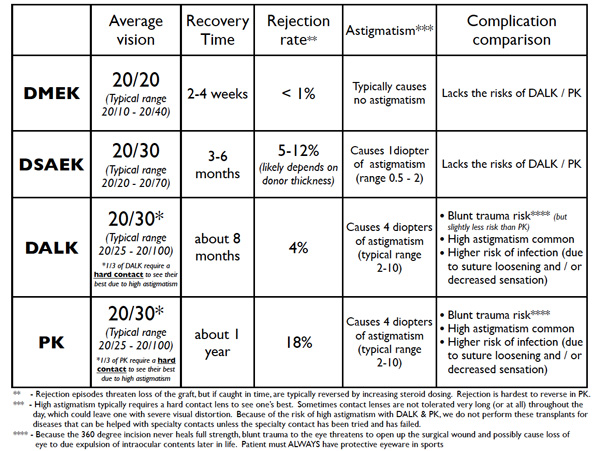
Not that long ago, the only available surgery was a full thickness cornea transplant or penetrating keratoplasty (PK). With PK, all of the corneal layers are transplanted, and new epithelial cells grow over the new cornea in time. The entire wall is knocked out with a hammer, and a new wall and all its layers are brought in. The owner provides a new layer of clear coat over time. A prosthetic corneal transplant (keratoprosthesis (KPro) is an artificial full-thickness corneal transplant.

Selective keratoplasty surgeries are a huge advance over PK in that they allow us to replace just the portion of the cornea that is diseased.

Selective Keratoplasty Diagram

    • Superficial keratectomy (SK) and phototherapeutic keratectomy (PTK) remove the epithelium (and possibly some superficial stroma with PTK) so that new healthier epithelium can grow in. The corneal epithelium is the only layer of the cornea that regenerates. So, SK and PTK aren’t true transplants. They are like removing some imperfections in the layer of clear coat on the outside of the house so that the owner can lay down new clear coat. Conversely, an epithelial stem cell transplant is necessary when the owner’s can of clear coat runs out or goes bad. It becomes necessary to give the owner a new supply of clear coat, which involves transplanting in a special area of a donor cornea.
    • Deep anterior lamellar keratoplasty (DALK) transplants all stroma but leaves the host’s healthy Descemet’s membrane and endothelium behind to allow less risk of rejection or less risk from blunt trauma. The entire wall is changed out, but the host’s own delicate wallpaper is left behind.
  • Descemet’s membrane endothelial keratoplasty (DMEK) and Descemet’s stripping automated endothelial keratoplasty (DSAEK) replace Descemet’s membrane and endothelium without removing the host’s stroma. DMEK is more selective than DSAEK. Both DMEK and DSAEK remove old Descemet’s membrane and endothelium. DMEK adds a new Descemet’s membrane and endothelium only. DSAEK also adds a new Descemet’s membrane and endothelium but with an additional layer of donor stroma. Using the wallpaper analogy, in both DMEK and DSAEK the old wallpaper is removed. With DMEK, only new wallpaper is inserted. With DSAEK, a new piece of drywall that has new wallpaper on it is inserted on top of the old drywall.

Why do we need different types of transplants? Why not just do PK for everyone

PK has many limitations regarding vision, astigmatism, recovery time, rejection rate, and other risks. All types of selective transplants improve the outcomes when compared to PK. DMEK especially has many other benefits compared to DSAEK and PK. For example, performing PK for purely endothelial disease is outdated. It would be like knocking down the wall and bringing in a whole new wall when the only thing needed is new wallpaper!

Cornea Transplant Procedures Diagram

Do I need to go on systemic immunosuppressive medications for a corneal transplant?

Not for most types of corneal transplants. Epithelial stem cell transplants are an exception. When someone gets a kidney or a heart transplant, they need to go on strong medications that make the immune system less aggressive and less likely to attack the transplant. These medications have some potentially significant side effects.

Because there are no blood vessels in the cornea, it is generally invisible to the immune system. Thus for corneal transplants, with the exception of epithelial stem cell transplants, we only need anti-inflammatory eye drops to prevent an immune system attack or rejection. Most patients get down to one drop a day after several few months.

Rarely, patients with aggressive immune systems require stronger systemic medications to prevent rejection. Even with aggressive immune systems, rejection is still very rare with DMEK and is more commonly seen with DSAEK, DALK, or PK. PK has the highest risk of rejection.

After a corneal transplant, do I have to take any precautions if I ever receive a vaccination?

There is some data to suggest that the increase in the immune system activity after vaccination, including the flu and shingles vaccines, may put the graft at a slightly increased risk for a rejection episode. To combat this, we typically recommend that if you were down to just one steroid drop a day, immediately following a vaccine you should increase the steroid drops to four times a day for one week, then two times a day for one week, and then go back to just one daily. If you have previously been told that you cannot take steroid drops due to pressure problems or other issues, ask your cornea surgeon for advice before getting a vaccine. transplant.

How long will my transplant last?

Traditional full thickness corneal transplants (PK) last about 20 years. Cell count studies show that, with the passage of time, transplants still lose endothelial cells gradually just like any other cornea, but usually at a faster rate. When the endothelial cell counts fall low enough, the transplant becomes opaque and fails. Since DMEK and DSAEK are relatively new, it is not possible to say how long they will last; however, preliminary data is encouraging, especially for DMEK. There is variation between transplants, but early data suggest some transplants can even last one’s lifetime. Either way, the replacement of a transplant is possible.

Dr. Tenkman is studying variables that may reveal which donors have cells that are more resistant to death and also surgical techniques that are minimally harmful to endothelial cells. Many surgeons suggest it is normal to lose 30 to 50% of the donor’s endothelial cells during surgery. We have some early data suggesting less than 10% cell loss from surgery when selecting a specific subset of donors.

Does a rejection episode mean that I will lose my cornea transplant?

No. If untreated, a rejection episode can cause significant damage. But usually, the episode stops with an increase in anti-inflammatory eye drops. Patients are instructed to RSVP: come see us if they develop Redness, Sensitivity to light, Vision reduction, or Pain. It’s key to see us right away for prompt diagnosis and treatment.

Will I need glasses after my transplant? Could laser vision correction be done to reduce any postoperative need for glasses?

Whether or not you need glasses after your transplant depends on many factors. DMEK transplants reduce glasses dependence the most often. DSAEK is less predictable. DALK and PK can both frequently cause high astigmatism that needs glasses or even hard contacts to attain good vision.

Some patients are candidates for ASA (advanced surface ablation) to minimize their dependence on glasses or contacts after corneal transplant surgery. ASA is a laser vision correction procedure similar to LASIK. Whether or not ASA could be done to reduce dependence on glasses or contacts after surgery depends on several factors and is taken on a case by case basis.

What are the costs and risks of a cornea transplant?

Corneal transplant tissue is donated, but still typically costs $3,700 or more. The fees go to the eye banks that must harvest the corneas, screen for possible diseases that could be transferred to the patient, transport, and process the corneas, and deliver them to the surgery center… all within just a few days. The cost of receiving a transplant includes not just the tissue, but the surgery center, surgeon, and anesthesia fees. Fortunately, these costs are less at an outpatient surgery center and are typically covered by Medicare and private insurance, although patients may be responsible for deductibles and co-payments. Generally, DMEK and DSAEK can be performed in an outpatient surgery center whereas DALK and PK more often have to be done at a hospital. Having surgery at a hospital can increase costs several-fold.

The risk of infection from a corneal transplantation procedure is about the same as following a routine blood transfusion. The risk of receiving a disease from a transplant is very low.

Other risks include primary graft failure, rejection, graft dislocation, cataract formation, glaucomainfection, irregular astigmatism, double vision, bleeding, iris damage, vitreous prolapse, and cystoid macular edema. Most of these complications are quite rare but are theoretically possible with all types of eye surgery. Patients using steroid drops, which all corneal transplants require, have to be followed for the development of increased pressure. High eye pressure usually gives no symptoms, so follow up is crucial to allow the doctor to treat as necessary with medication or procedures. Patients are usually seen every 3 to 6 months for life depending on the strength of steroid used.

In our hands, the overall success rate for a DMEK or DSAEK graft attaching and working well is about 99%. Should the transplant fail due to rejection or otherwise, it would need to be repeated.

Because the time to heal is longer for DALK and PK, and because they involve more risk, the odds of a DALK or PK failing to restore vision is more significant than for DMEK or DSAEK. DALK and PK have several additional risks.

  • DALK, and more so PK, have a higher risk of bleeding during surgery while the patient’s own cornea is removed and the eye is depressurized. Although not common, such bleeding can do significant damage to the eye.
  • DALK, and more so PK, also have a significant risk of rejection. Higher doses of steroid drops may be necessary in here to prevent or treat rejection, which can increase the risk of increased eye pressure (glaucoma).
  • DALK and PK involve a 360-degree corneal incision. Such large corneal wounds don’t heal with full strength and are at risk to split open with moderate blunt trauma (ie if a patient fell and hit their face in the shower). If the wound were to split open, it could lead to loss of the eye.
  • DALK and PK require about 16 sutures. These sutures are not removed for many months or even over a year. The sutures are buried in a manner so they cause no pain to the patient. However, they commonly become loose and can cause a foreign body or sandy sensation. More significantly, a loose suture is a risk for infection (corneal ulcer). Corneal ulcers can be more serious in the setting of a corneal transplant because the immune system in the area is suppressed by steroid eye drops.
  • DALK and PK commonly heal with large amounts of astigmatism. The normally spherical corneal surface becomes like an irregularly shaped egg. Since the smooth corneal shape largely determines focus, an irregular cornea causes blurred vision. Corneal astigmatism is measured in diopters. In routine patients who have not had had a corneal transplant, we consider 1 diopter mild astigmatism, 2 diopters moderate astigmatism, and 3 diopters high astigmatism. DALK and PK average about 4 to 5 diopters of astigmatism. A common range is 2 to 10 diopters of astigmatism. Regarding astigmatism risk, there are controllable and uncontrollable causes. The surgeon can try to sew the graft as evenly as possible to reduce risk of severe astigmatism; however, the surgeon cannot control the natural tension lines in the cornea or how symmetrically the tension lines will balance as the wound heals. Severe or irregular astigmatism cannot be corrected with glasses. Special hard contact lenses are often necessary. On average, one-third of all DALK and PK patients need hard contact to see their best due to astigmatism. Therefore, at Bennett & Bloom, we never perform DALK or PK for keratoconus patients to try to get out of contact lenses. The patient could go through all the added cost and risk of surgery only to end up where they started… or worse. DALK and PK are reserved for severe disease that cannot be fixed by contact lens wear


For corneal transplant at Bennet & Bloom Eye center in the USA or for medical tourism in the USA, contact Eyeupdate Clinic & Optical Supplies, 01 Ajuwon junction, Off Elliot bus stop, Iju Ishagah, Lagos.Tel: +2347030000001, +19093663551

Muscles, Nerves, and Blood Vessels in the Human Eye

By David Terfera, Shereen Jegtvig

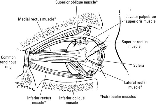
Muscles enable you to move your eyes. Ocular nerves allow you to interpret what you see and blood vessels keep your eyes oxygenated. Six muscles, collectively called the extraocular muscles, move the eyeball. A seventh muscle moves the eyelid and is also found in the orbit.

The muscles of the human eye

The following muscles help your eyes move around.

image0.jpg

  • Levator palpebrae superioris: Originates on the sphenoid bone above the optic canal. It inserts into the superior tarsis and skin of the eyelid. It’s innervated by the oculomotor nerve and elevates the superior eyelid.

  • Superior oblique: Originates on the sphenoid bone and inserts into the sclera deep to the superior rectus muscle. It’s innervated by the trochlear nerve and abducts, depresses, and medially rotates the eyeball.

  • Inferior oblique: Originates on the anterior part of the orbital floor and inserts onto the sclera deep to the lateral rectus muscle. It’s innervated by the oculomotor nerve and abducts, elevates, and laterally rotates the eyeball.

  • Superior rectus: Originates on the common tendinous ring and inserts into the sclera behind the corneoscleral junction. It’s innervated by the oculomotor nerve, and it elevates, adducts, and medially rotates the eyeball.

  • Inferior rectus: Originates on the common tendinous ring and inserts into the sclera behind the corneoscleral junction. It’s innervated by the oculomotor nerve and depresses, adducts, and laterally rotates the eyeball.

  • Medial rectus: Originates on the common tendinous ring and inserts into the sclera behind the corneoscleral junction, this muscle is innervated by the oculomotor nerve and adducts the eyeball.

  • Lateral rectus: Originates on the common tendinous ring and inserts into the sclera behind the corneoscleral junction. It’s innervated by the abducent nerve and abducts the eyeball.

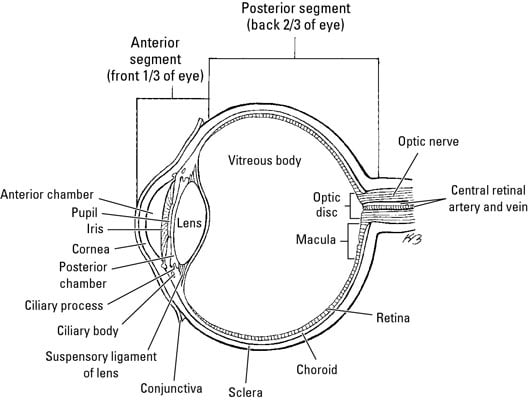
The nerves of the eye

The eyes are served by the following cranial nerves and their branches:

image1.jpg

  • Optic nerve (CN II): Sensory nerve that transmits impulses from the retina to the brain

    • Oculomotor nerve (CN III), trochlear nerve (CN IV), and abducent nerve (CN VI): Enter the orbital space through the superior orbital fissure to innervate the extraocular muscles.

    • Ophthalmic nerve (part of the trigeminal nerve, CN V): This nerve has three branches:

      • The lacrimal nerve runs to the lacrimal gland and gives off branches to the conjunctiva and skin of the superior eyelid.

      • The frontal nerve enters through the superior orbital fissure and provides sensory innervation to the superior eyelid, scalp, and forehead.

      • The nasociliary nerve is the sensory nerve to the eyeball. It also has branches that serve the orbit and other parts of the face. One of its branches, the infratrochlear nerve, supplies the eyelids, conjunctiva, and lacrimal sac.

    • Ciliary ganglion: This group of postsynaptic parasympathetic nerve cell bodies is associated with the oculomotor nerve and ophthalmic nerve (CN V1). Presynaptic parasympathetic fibers from the oculomotor nerve synapse on the cell bodies of postsynaptic parasympathetic neurons in the ciliary ganglion.

      Short ciliary nerves emerge from the ciliary ganglion and enter the eye. The short ciliary nerves contain postsynaptic parasympathetic fibers from the ciliary ganglion, afferent fibers of the nasociliary nerve, and postsynaptic sympathetic fibers from the internal carotid plexus. Postsynaptic parasympathetic fibers innervate the ciliary muscle and sphincter pupillae muscle. Afferent fibers convey sensory impulses from the iris and cornea. Postsynaptic sympathetic fibers innervate the dilator pupillae muscle.

      The long ciliary nerves contain afferent and postsynaptic sympathetic fibers from the nasociliary nerve. Long ciliary nerves bypass the ciliary ganglion and run to the iris, cornea, and dilator pupillae muscle.

    The blood vessels

    Blood flow to the orbit (and beyond) comes from branches of the internal carotid artery, chiefly via the ophthalmic artery and its branches:

    • Ophthalmic artery: Branches from the internal carotid artery and passes through the optic canal into the orbital cavity

    • Central artery of the retina: Runs from the ophthalmic artery to the eyeball alongside the optic nerve; it branches at the optic disc and supplies the retina

    • Supraorbital artery: Starts at the ophthalmic artery and exits the orbit at the supraorbital notch to supply the forehead and scalp

    • Supratrochlear artery: Runs from the ophthalmic artery to the forehead and scalp

    • Lacrimal artery: Runs from the ophthalmic artery along the lateral rectus muscle to supply the lacrimal gland, conjunctiva, and the eyelids

    • Dorsal nasal artery: Branches from the ophthalmic artery and runs along the nose to supply it with blood

    • Short posterior ciliary arteries: Branch from the ophthalmic artery and pierce the sclera at the edge of the optic nerve; they supply the choroid and the rods and cones of the retina

    • Long posterior ciliary arteries: Branch from the ophthalmic artery and pierce the sclera to supply the ciliary body and iris

    • Posterior ethmoidal artery: Leaves the ophthalmic artery to supply blood to ethmoidal cells

    • Anterior ethmoidal artery: Runs from the ophthalmic artery to supply ethmoidal cells, frontal sinus, nasal cavity, and skin over the nose

    • Anterior ciliary artery: Runs from the muscular branches of the ophthalmic artery through the sclera near the rectus muscles and forms an arterial network in the iris and ciliary body

    • Infraorbital artery: Runs from the maxillary artery along the infraorbital groove and out to the face

    Blood is returned from the orbits via the superior and inferior ophthalmic veins, which pass through the superior orbital fissure into the cavernous sinus. The central vein of the retina may join an ophthalmic vein or enter the cavernous sinus directly. Vorticose veins drain the vascular layer of the eyeball, and the scleral venous sinus encircles the anterior chamber of the eyeball.

How alcohol affects your eyes -Eye care perspective

Although light consumption of alcohol probably won’t cause any health problems, drinking alcohol excessively can have harmful effects on your body, including your eyes. Heavy drinking of alcohol may cause problems with your vision and overall eye health including the following:

    • Decreased visual performance: Your overall visual performance may be altered since drinking heavily impairs brain function. You may have blurred vision or double vision due to weakened eye muscle coordination. You may also experience delayed reactions while driving.
    • Slow pupil reactions: Alcohol tends to affect the speed at which your iris constricts and dilates. A driver that has been drinking alcohol cannot adapt as quickly to oncoming headlights.
    • Decreased peripheral vision: Drinking alcohol has also been shown to decrease the sensitivity of your peripheral vision. This may give you the effect or perception of having tunnel vision.
  • Decreased contrast sensitivity: Drinking too much alcohol can alter your contrast sensitivity, or how precise you can discern between shades of gray. Driving in rain or fog will be much more dangerous.
  • Optic neuropathy: Also referred to as tobacco-alcohol amblyopia, people who drink or smoke in excess can develop optic neuropathy. You might develop a painless loss of vision, decreased peripheral vision or reduced color vision. Even though studies have shown the vision loss to be a result of a nutritional deficiency, some professionals believe that the condition develops because of toxic effects of alcohol and tobacco.
  • Frequent migraines: Alcohol has been shown to be a trigger for severe migraine headaches in some people. You may experience a temporary, but debilitating visual aura before the onset of the headache. The visual aura may appear as blind spots, graying of vision or zig-zag patterns of light.​
  • Poor cosmetic appearance: Drinking can cause eye redness. Alcohol causes the blood vessels in your eyes to expand, making them more prominent.


If you have any issues with your eyes, visit or call : Eye update Eye clinic & optical supplies, 01, Ajuwon junction, Ajuwon bus stop, Akute/Ajuwon road, off Elliot bus stop, Iju-Ishagah. Tel: 08034971582

Postal Codes of Streets in Ojodu Lagos Nigeria

Ojodu Streets Zip Codes

Address:
Area: Ojodu, Lagos, Lagos State. Associated Zip Code: 100213

District Name: Area: Ojodu
Local Government Area: Lagos
State: Lagos State
Associated Zip Code: 100213

Streets: (260)
Street name Zip Code
Raimi St St. 100213
Rasheed Baruwa St. 100213
Regina Ola Ore St St. 100213
Sogo St St. 100213
Sowebo Oladipupo St. 100213
Soyemi St St. 100213
Sule Abore St St. 100213
Taiwo Ishola St. 100213
Taiwo Okolu St. 100213
Tijani Bello St St. 100213
Unity Cl St. 100213
Zinzon S St. 100213
Alara St St. 100213
Adesina St St. 100213
Funsho St St. 100213
Tobun St St. 100213
Olumo St St. 100213
OlasimboSt St. 100213
Olu Osifeso St St. 100213
Remi Abuah St St. 100213
Majaro St St. 100213
Akintunde St St. 100213
Mobolaji St St. 100213
Johnson St St. 100213
Araromi St St. 100213
Ajayi St St. 100213
Aderibigbe St St. 100213
Lawal St St. 100213
Adebiyi St St. 100213
Abiodun St St. 100213
Adesina St St. 100213
Barikisu Iyade St St. 100213
University Rd St. 100213
Davies St St. 100213
Ayodele St St. 100213
Amusa St St. 100213
Sule St St. 100213
Abudu St St. 100213
Amen St St. 100213
Ajayi Bembe St St. 100213
Obadina St St. 100213
Mosuro St St. 100213
Sadiku St St. 100213
Ebun St St. 100213
Balogun St St. 100213
Are Ago St St. 100213
Abule Ijesha Rd St. 100213
Morris Rd St. 100213
Eletu Odibo St St. 100213
Shodipe St St. 100213
Saka Ln St. 100213
Martins St St. 100213
Odenike St St. 100213
Shobowale St St. 100213
Otun St St. 100213
Oduntan St St. 100213
Oguntuga St St. 100213
Silver St St. 100213
Bailey St St. 100213
Akinsola St St. 100213
Bamigbopa St St. 100213
Alaka St St. 100213
Mosuro St St. 100213
Bankole St St. 100213
Lemboye St St. 100213
Ogabi St St. 100213
Iwaya Rd St. 100213
Owodunni St St. 100213
Memuda St St. 100213
Ogunkoya St St. 100213
Abiye St St. 100213
Ijebu Quarters St. 100213
Oloto St St. 100213
Victoria St St. 100213
Ajoke St St. 100213
Aderupoko St St. 100213
Imoru St St. 100213
Audu Bale St St. 100213
Adegbenro St St. 100213
Salami St St. 100213
Church St St. 100213
Akanni St St. 100213
Omitogun St St. 100213
Yey St St. 100213
Ogunbiyi St St. 100213
Omotola St St. 100213
Arowolo St St. 100213
Balogun St St. 100213
Moshalashi St St. 100213
Akintola St St. 100213
Owode St St. 100213
Pedro St St. 100213
Omiaro St St. 100213
Ogbwe St St. 100213
Oyewale St St. 100213
Onituere St St. 100213
Ogundimu St St. 100213
Joseph Marrison St St. 100213
Gbede St St. 100213
Dacosta St St. 100213
Ogayemi St St. 100213
Afolabi Ayorinde St St. 100213
Wugbo St St. 100213
Yovi St St. 100213
Akinlehinb St St. 100213
Ayato St St. 100213
Oke Anaiye St St. 100213
Igbore St St. 100213
Development St St. 100213
ALh. Rainmi St. 100213
Araromi St St. 100213
Iman Ln St. 100213
Wright St St. 100213
Makoko Rd St. 100213
Dany Est St. 100213
Akinbo St St. 100213
Erejuwa St St. 100213
Ramotu St St. 100213
Olaiya St St. 100213
Church St St. 100213
Ajelekoko St St. 100213
Igbehin Adun St St. 100213
Abudu St St. 100213
Rafiu St St. 100213
Adetia St St. 100213
Olulu St St. 100213
Olumide St St. 100213
Sariyu St St. 100213
Obalomu St St. 100213
Falodun St St. 100213
Olayide St St. 100213
Alafia St St. 100213
Oluwatoyin St St. 100213
Bale St St. 100213
Alh.Sule St St. 100213
Alh. Raimi St. 100213
St Finbar College St St. 100213
Moronfolu St St. 100213
Community Rd St. 100213
Obayan St St. 100213
Amos Bakare St St. 100213
Afolabi Brown St St. 100213
Funmilayo Oyaronke St St. 100213
Tunde Bello St St. 100213
Emily Akinola St St. 100213
Iyiola Olawale St St. 100213
AdeyinkaOsijo St St. 100213
Abdulahi St St. 100213
Wulemotu Ajoke St St. 100213
Abayomi St St. 100213
Olarewaju St St. 100213
Rikett Cl St. 100213
Ayetoro St St. 100213
Oyenuga St St. 100213
Oridami St St. 100213
Itesiwaju St St. 100213
Sholanke St St. 100213
Shuwu Court St. 100213
Mafowoku St St. 100213
Adenuga Kajaro St St. 100213
Ojo St St. 100213
Abiodun Cl St. 100213
Bandi Adewale St St. 100213
Olorunjare St St. 100213
Ayinde St St. 100213
Onafeko St St. 100213
Adetayo Osho St St. 100213
Osanyinpeju St St. 100213
Abatan Rd Rd. 100213
Alonge St St. 100213
Abigi St St. 100213
Awolaja St St. 100213
Abiodun St St. 100213
Oluwole St St. 100213
Adebisi Awosoga 100213
Animashaun St St. 100213
Adebolawale St St. 100213
Adegoke Ajayi St St. 100213
Ademola Ajasa 100213
Adeniyi Oke St St. 100213
Adewale 100213
Adewale Adegun 100213
Adeyemo Akapo 100213
Adisa Ambali Cl Cl. 100213
Agoro 100213
Aina St St. 100213
Ajana Cl Cl. 100213
Ajayi St St. 100213
Akin Oladeguro 100213
Akinola St St. 100213
Akinsanya St. 100213
Akiode Cr St. 100213
Alh. Abass St St. 100213
Alh. Ashafa St St. 100213
Alh.Bakare St. 100213
Alh. Bashiru St St. 100213
Ali Baogun St. 100213
Animashaun St St. 100213
Anu Oluwapo St St. 100213
Ayo Alabi St St. 100213
Babatunde Ladega St. 100213
Bakare St St. 100213
Bamako St St. 100213
Bank Shonibare St St. 100213
Banni St St. 100213
Basiaru St St. 100213
Beatrice Spencer St. 100213
Bisi Gbadina St St. 100213
Bosude St St. 100213
Bunuyo Cl St. 100213
Burdland St St. 100213
College Rd St. 100213
Arogangan St. 100213
Debo Aina St. 100213
Efon Alaye St. 100213
Foly St St. 100213
Gbenga Kilo St St. 100213
Gbite St St. 100213
Gbolahon Owolabi Cl St. 100213
Ibadan Ewy St. 100213
Ibrahim Adekunle St St. 100213
Ifo Cl St. 100213
Irepodun St St. 100213
Isale Tapa St St. 100213
Isheri Holiday Inn St St. 100213
Isheri Rd St. 100213
Ishola Bello St St. 100213
Ivern Ave St. 100213
Jacob Runsance Cl St. 100213
Jide Ayo St. 100213
Kadiri St. 100213
Kayode Ali Cl St. 100213
Kupoluyi St St. 100213
LSDPC Quarters St. 100213
Lola Holloway St. 100213
Mafaosky Cl St. 100213
Moses Adebayo St St. 100213
Muyibo St St. 100213
Muyiwa Oyefusi St. 100213
New Isheri Rd St. 100213
Oba Bobington Ashoye St. 100213
Obafolabi St St. 100213
Oboku St St. 100213
Odozi St St. 100213
Ogunbiyi Akande St. 100213
Ola Oluwa St St. 100213
Oladipupo Oluwole St St. 100213
Olajide Ave St. 100213
Olaleke Taiwo St St. 100213
Olanipekun St. 100213
Olatunde Badmus St. 100213
Olawale St. 100213
Olumuyiwa St. 100213
Oluwadamilola Falade St. 100213
Oluwatoyin Cl St. 100213
Omofade Cr St. 100213
Omorogie St St. 100213
Orimeta St St. 100213
Popoola Oyinloye Cl St. 100213
Popola St St. 100213