How to detect early signs of heart disease in the eyes

Early Signs of Heart Diseases Appear in the Eyes

According to experts, Eye doctors may be able to detect signs of heart disease during a comprehensive eye exam. A new study finds that people with heart disease tend to have retinas marked by evidence of eye stroke.

Eye strokes happen when the eye is deprived of blood flow and oxygen, causing cells to die. This creates a mark, called a retinal ischemic perivascular lesion. These marks can be spotted when eye doctors run optical coherence tomography, or OCT is order to take a closer look at the retina.

OCT scans of the retina are valuable ways to detect disease and dysfunction in all parts of the body — not just the eyes. Eye scans can detect signs of Alzheimer’s, Parkinson’s and other underlying health conditions.

How eye exams can detect heart disease

The eye is the only place in the body where a doctor can see the live action of blood vessels, nerves and connecting tissue without relying on an invasive procedure. That’s why eye doctors are often the first to detect health conditions including high blood pressure, high cholesterol, stroke and more.

While the marks left behind by eye strokes may be present in low numbers in healthy people, those with heart disease tend to have a far greater number. Researchers arrived at these results by reviewing the medical records of 84 people with heart disease and 76 healthy people, all of whom had received a retinal OCT scan.

Muscles, Nerves, and Blood Vessels in the Human Eye

By David Terfera, Shereen Jegtvig

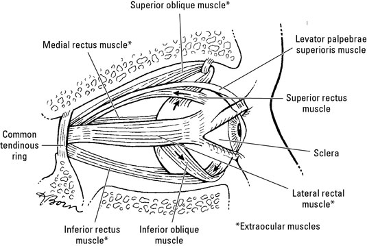
Muscles enable you to move your eyes. Ocular nerves allow you to interpret what you see and blood vessels keep your eyes oxygenated. Six muscles, collectively called the extraocular muscles, move the eyeball. A seventh muscle moves the eyelid and is also found in the orbit.

The muscles of the human eye

The following muscles help your eyes move around.

image0.jpg

  • Levator palpebrae superioris: Originates on the sphenoid bone above the optic canal. It inserts into the superior tarsis and skin of the eyelid. It’s innervated by the oculomotor nerve and elevates the superior eyelid.

  • Superior oblique: Originates on the sphenoid bone and inserts into the sclera deep to the superior rectus muscle. It’s innervated by the trochlear nerve and abducts, depresses, and medially rotates the eyeball.

  • Inferior oblique: Originates on the anterior part of the orbital floor and inserts onto the sclera deep to the lateral rectus muscle. It’s innervated by the oculomotor nerve and abducts, elevates, and laterally rotates the eyeball.

  • Superior rectus: Originates on the common tendinous ring and inserts into the sclera behind the corneoscleral junction. It’s innervated by the oculomotor nerve, and it elevates, adducts, and medially rotates the eyeball.

  • Inferior rectus: Originates on the common tendinous ring and inserts into the sclera behind the corneoscleral junction. It’s innervated by the oculomotor nerve and depresses, adducts, and laterally rotates the eyeball.

  • Medial rectus: Originates on the common tendinous ring and inserts into the sclera behind the corneoscleral junction, this muscle is innervated by the oculomotor nerve and adducts the eyeball.

  • Lateral rectus: Originates on the common tendinous ring and inserts into the sclera behind the corneoscleral junction. It’s innervated by the abducent nerve and abducts the eyeball.

The nerves of the eye

The eyes are served by the following cranial nerves and their branches:

image1.jpg

  • Optic nerve (CN II): Sensory nerve that transmits impulses from the retina to the brain

    • Oculomotor nerve (CN III), trochlear nerve (CN IV), and abducent nerve (CN VI): Enter the orbital space through the superior orbital fissure to innervate the extraocular muscles.

    • Ophthalmic nerve (part of the trigeminal nerve, CN V): This nerve has three branches:

      • The lacrimal nerve runs to the lacrimal gland and gives off branches to the conjunctiva and skin of the superior eyelid.

      • The frontal nerve enters through the superior orbital fissure and provides sensory innervation to the superior eyelid, scalp, and forehead.

      • The nasociliary nerve is the sensory nerve to the eyeball. It also has branches that serve the orbit and other parts of the face. One of its branches, the infratrochlear nerve, supplies the eyelids, conjunctiva, and lacrimal sac.

    • Ciliary ganglion: This group of postsynaptic parasympathetic nerve cell bodies is associated with the oculomotor nerve and ophthalmic nerve (CN V1). Presynaptic parasympathetic fibers from the oculomotor nerve synapse on the cell bodies of postsynaptic parasympathetic neurons in the ciliary ganglion.

      Short ciliary nerves emerge from the ciliary ganglion and enter the eye. The short ciliary nerves contain postsynaptic parasympathetic fibers from the ciliary ganglion, afferent fibers of the nasociliary nerve, and postsynaptic sympathetic fibers from the internal carotid plexus. Postsynaptic parasympathetic fibers innervate the ciliary muscle and sphincter pupillae muscle. Afferent fibers convey sensory impulses from the iris and cornea. Postsynaptic sympathetic fibers innervate the dilator pupillae muscle.

      The long ciliary nerves contain afferent and postsynaptic sympathetic fibers from the nasociliary nerve. Long ciliary nerves bypass the ciliary ganglion and run to the iris, cornea, and dilator pupillae muscle.

    The blood vessels

    Blood flow to the orbit (and beyond) comes from branches of the internal carotid artery, chiefly via the ophthalmic artery and its branches:

    • Ophthalmic artery: Branches from the internal carotid artery and passes through the optic canal into the orbital cavity

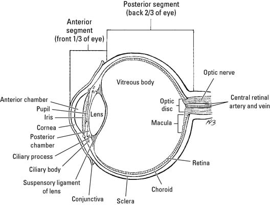
    • Central artery of the retina: Runs from the ophthalmic artery to the eyeball alongside the optic nerve; it branches at the optic disc and supplies the retina

    • Supraorbital artery: Starts at the ophthalmic artery and exits the orbit at the supraorbital notch to supply the forehead and scalp

    • Supratrochlear artery: Runs from the ophthalmic artery to the forehead and scalp

    • Lacrimal artery: Runs from the ophthalmic artery along the lateral rectus muscle to supply the lacrimal gland, conjunctiva, and the eyelids

    • Dorsal nasal artery: Branches from the ophthalmic artery and runs along the nose to supply it with blood

    • Short posterior ciliary arteries: Branch from the ophthalmic artery and pierce the sclera at the edge of the optic nerve; they supply the choroid and the rods and cones of the retina

    • Long posterior ciliary arteries: Branch from the ophthalmic artery and pierce the sclera to supply the ciliary body and iris

    • Posterior ethmoidal artery: Leaves the ophthalmic artery to supply blood to ethmoidal cells

    • Anterior ethmoidal artery: Runs from the ophthalmic artery to supply ethmoidal cells, frontal sinus, nasal cavity, and skin over the nose

    • Anterior ciliary artery: Runs from the muscular branches of the ophthalmic artery through the sclera near the rectus muscles and forms an arterial network in the iris and ciliary body

    • Infraorbital artery: Runs from the maxillary artery along the infraorbital groove and out to the face

    Blood is returned from the orbits via the superior and inferior ophthalmic veins, which pass through the superior orbital fissure into the cavernous sinus. The central vein of the retina may join an ophthalmic vein or enter the cavernous sinus directly. Vorticose veins drain the vascular layer of the eyeball, and the scleral venous sinus encircles the anterior chamber of the eyeball.